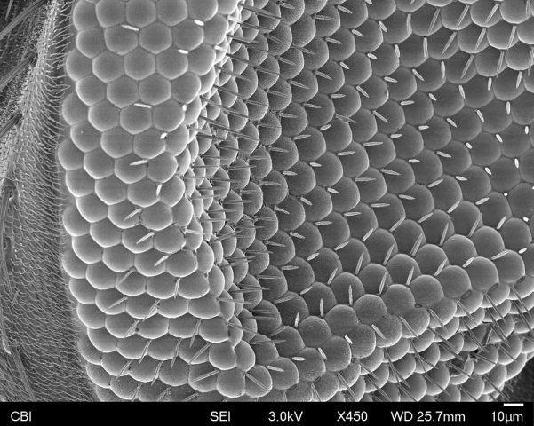
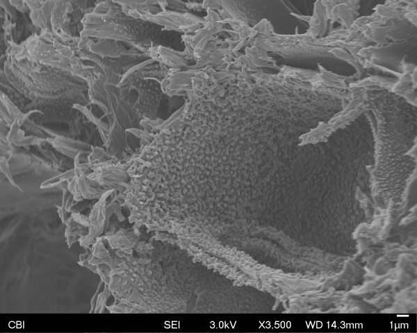
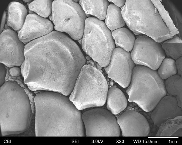
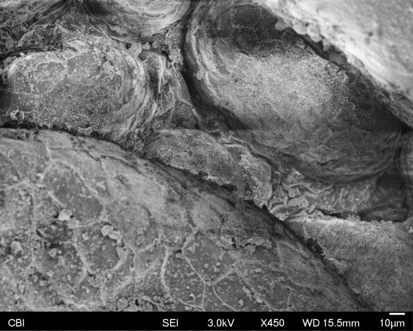
Compound eye of a Drosophila fruit-fly:
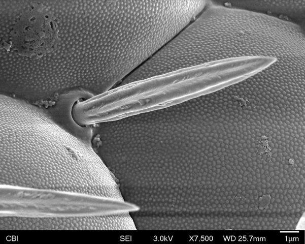
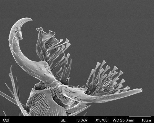
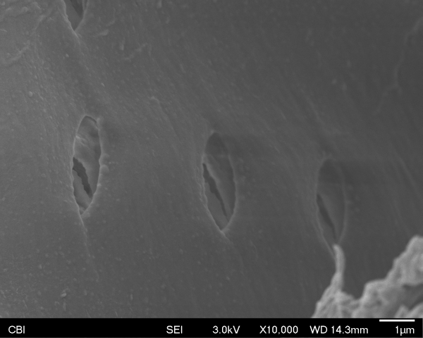
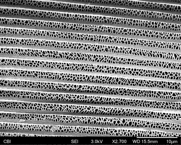
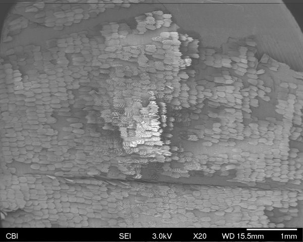
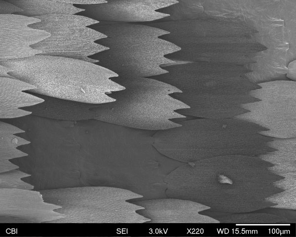
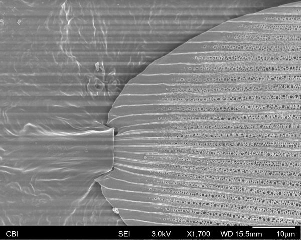
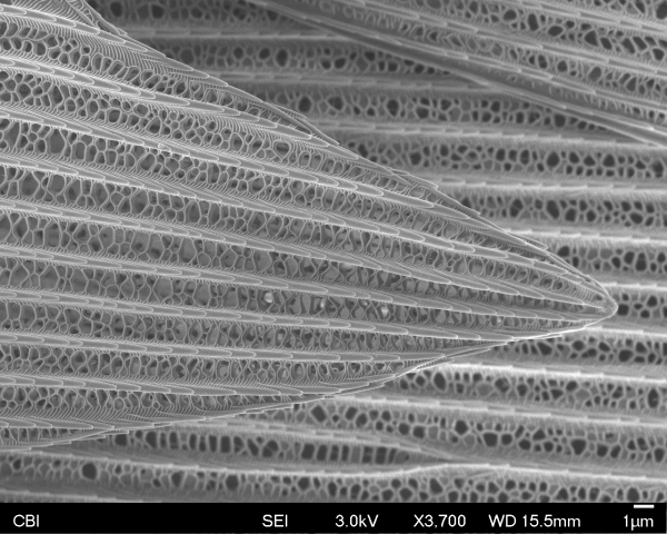
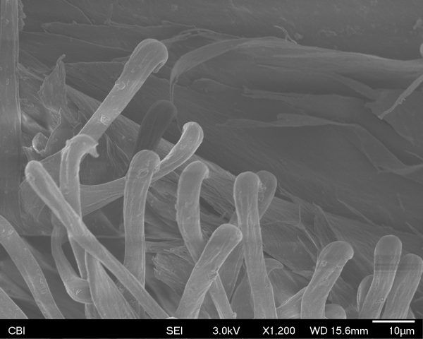
Foot of a fruit-fly:
Experimental Capture – Spring 2017
CMU School of Art, Spring 2017 • Prof. Golan Levin
Compound eye of a Drosophila fruit-fly:
Foot of a fruit-fly:
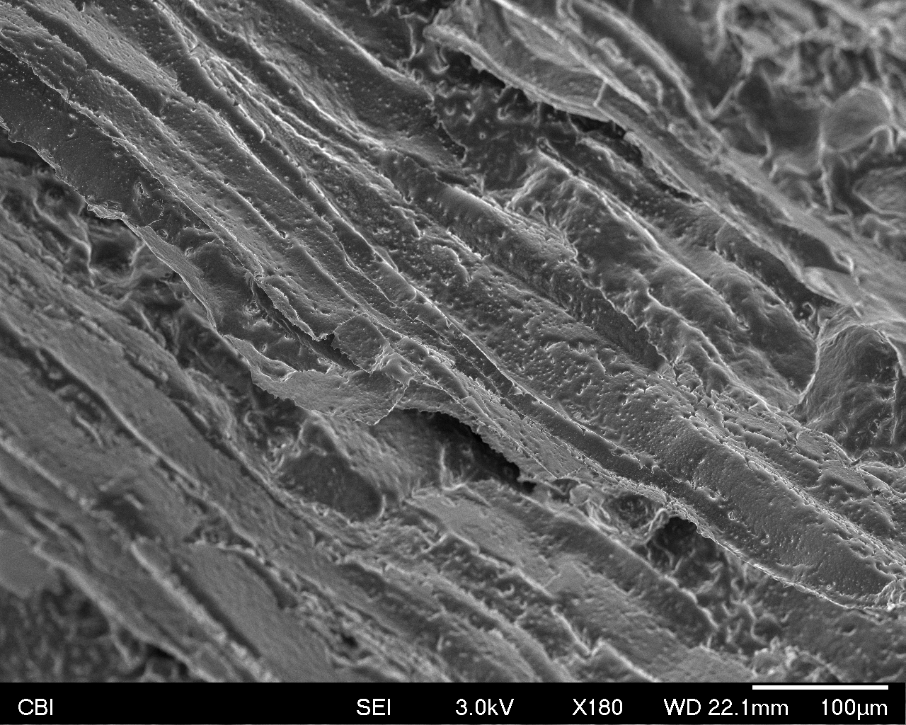
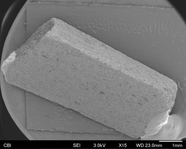
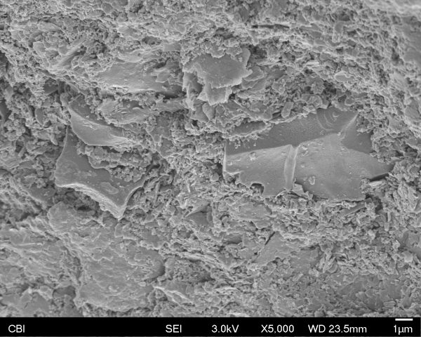
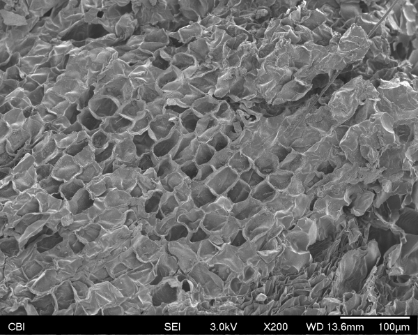
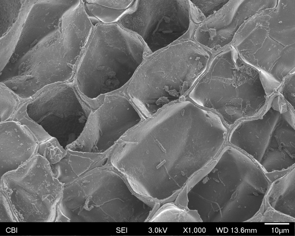
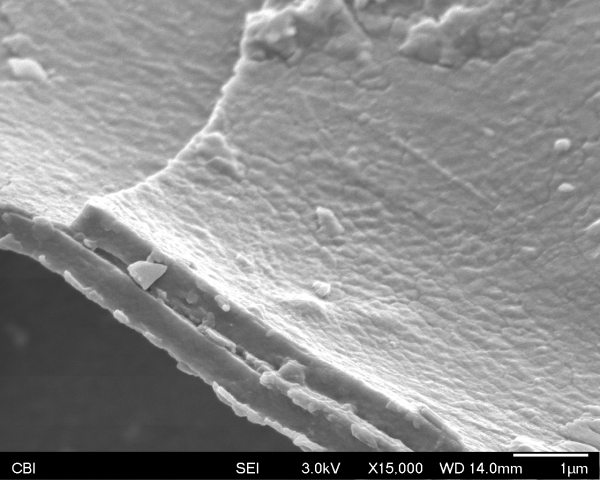
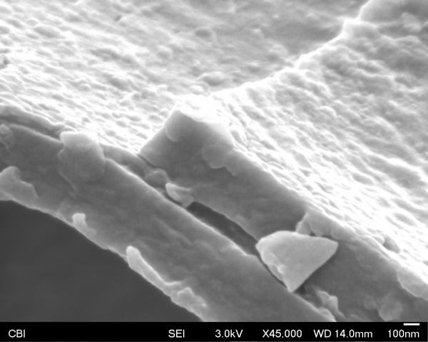
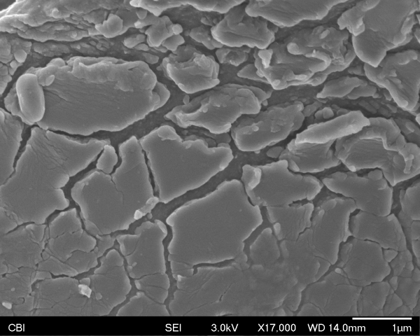
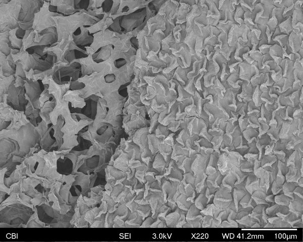
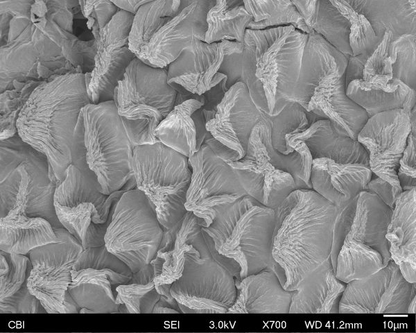
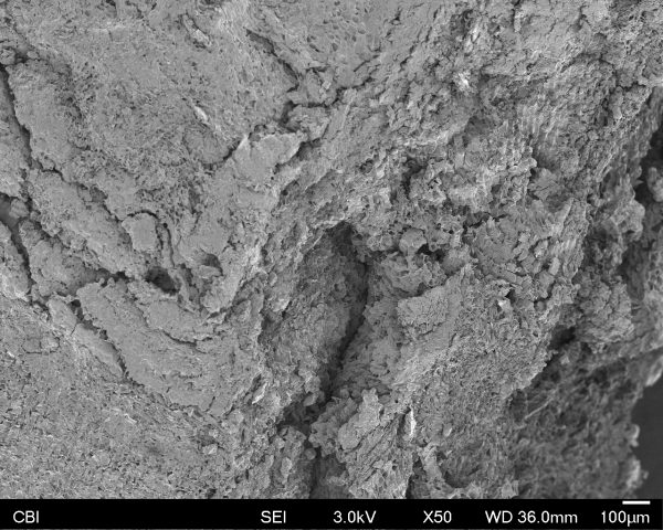
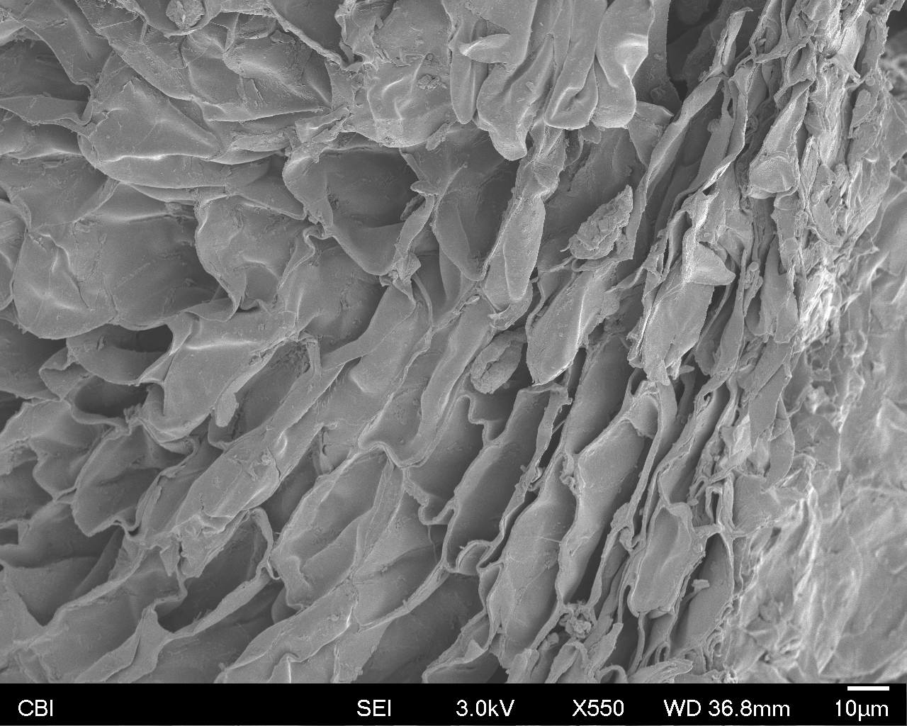
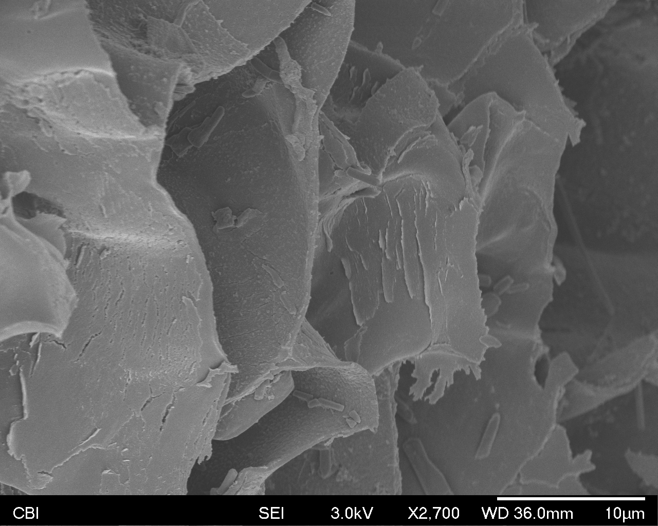
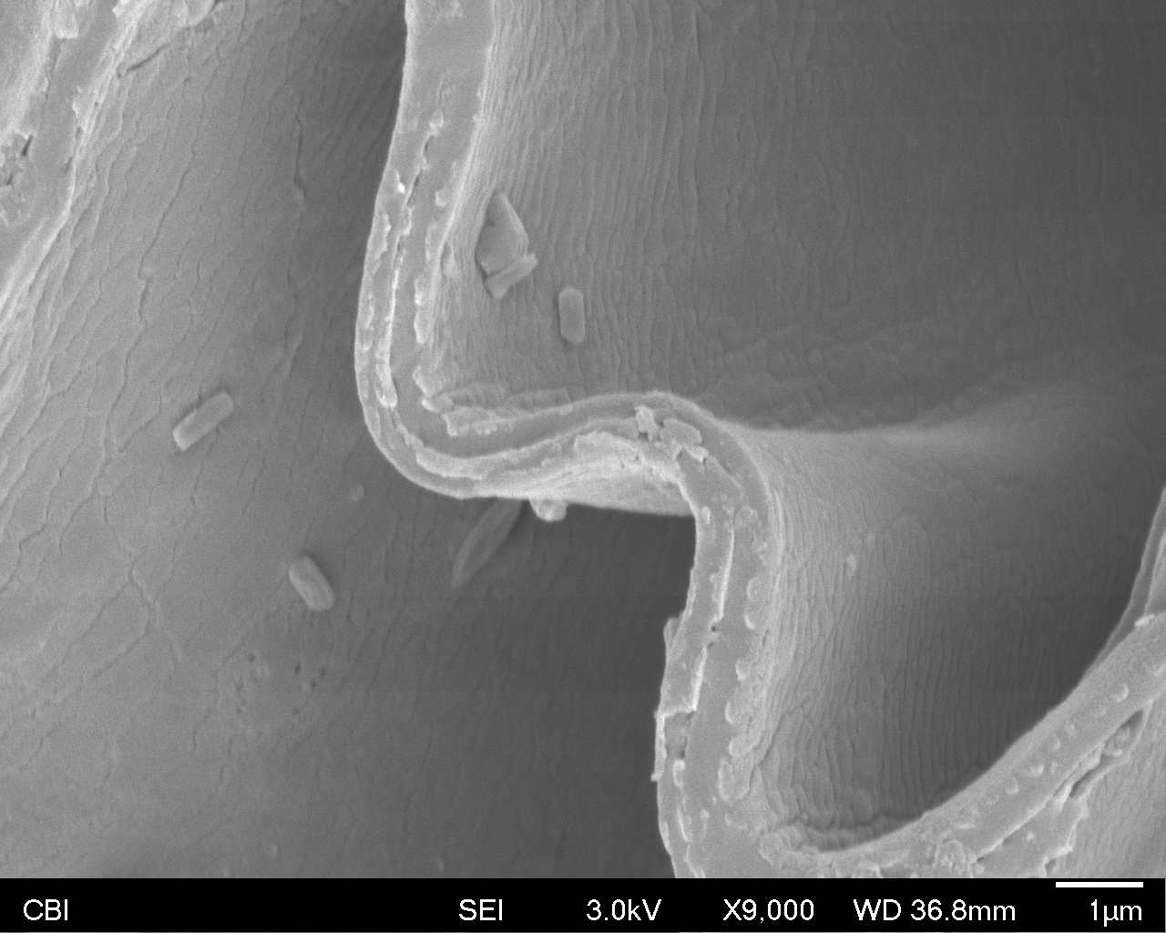
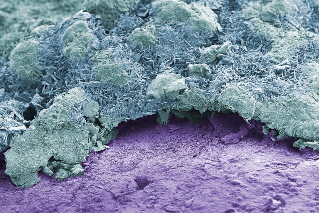
Lunch is Sandwich is Bacon
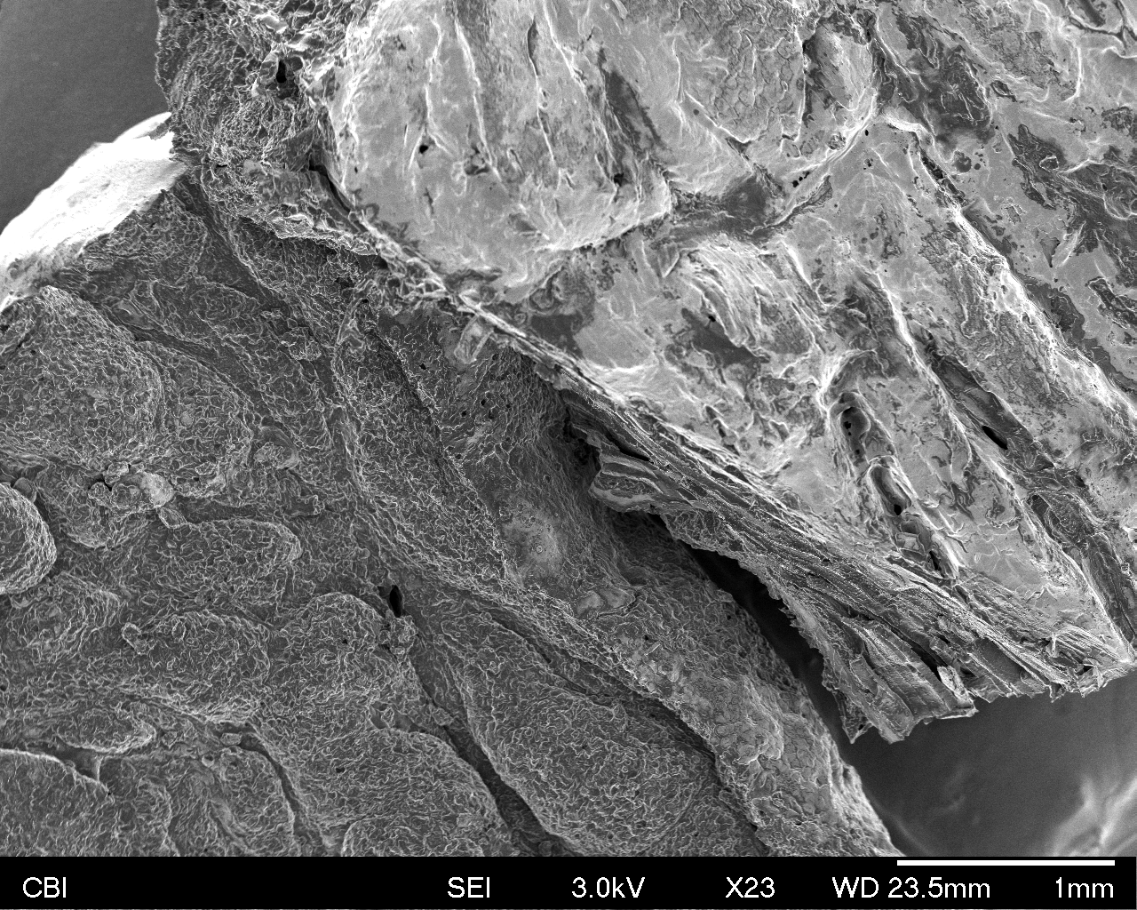
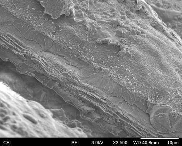
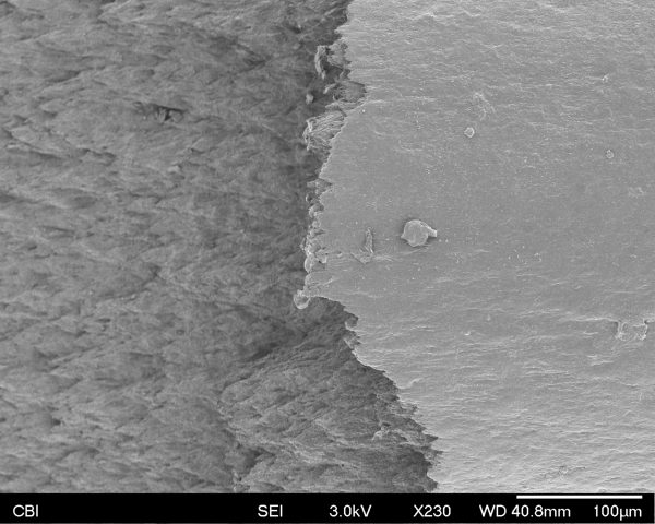
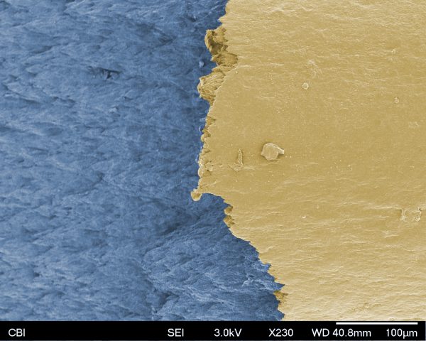
above: at left, the fatty tissue, at right, the muscle tissue
I really wanted to see what a smooth and transparent object would look under the SEM. After initially viewing my sample under the SEM, even though there was the expectation of seeing Something, I was still surprised by how much variation was present on the smooth plastic surface. The most interesting phenomenon I was multitudes of small cylindrical nubs sticking out of the surface, at the 500nm scale. I wonder how they interact with light and affect the appearance of the googly eye, as well as how they originate. In addition, in the final zoom-out, I was intrigued by the possibilities for creating abstract compositions using micrography.
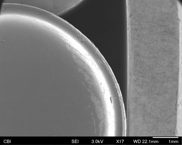
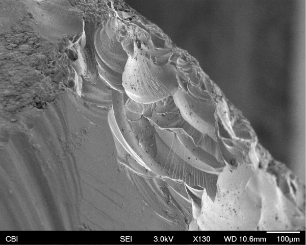
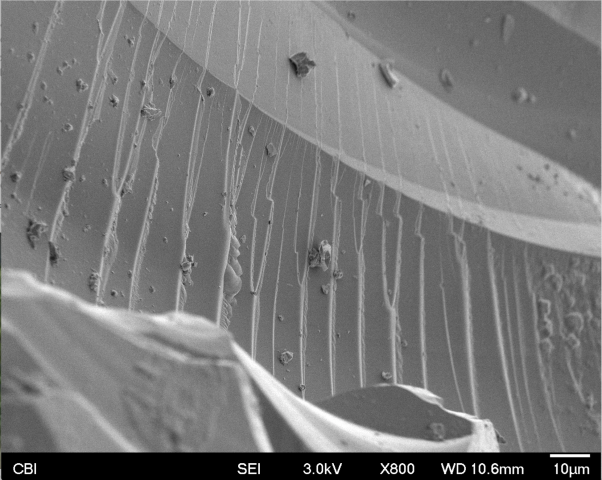
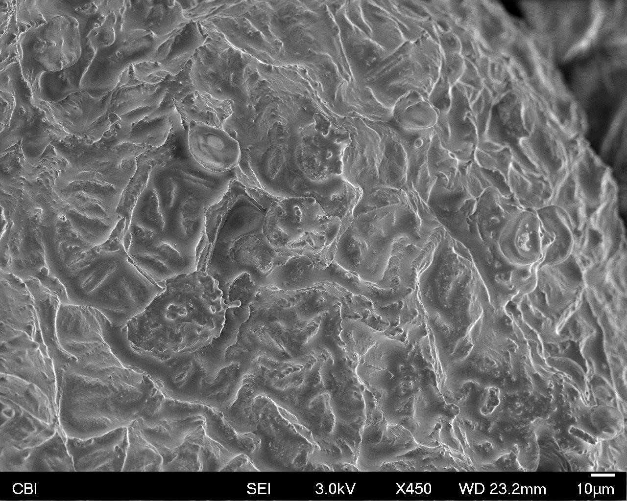
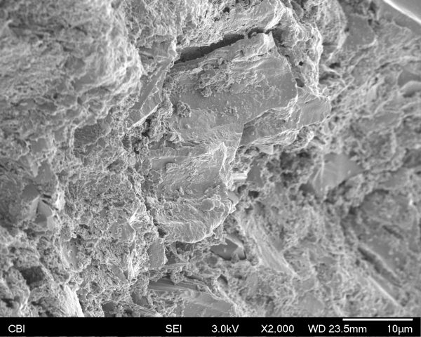
I decided to scan a small piece of frit (glass shard). The SEM machine was malfunctioning (joystick not working to move around the piece) while it was my turn, but I still got a few interesting images out of the process. I feel like I learned more about what a SEM machine is and also just how cool things look when you zoom in close. Even the most mundane objects look fascinating. I really enjoyed looking at the stress marks where the glass was broken. It gives almost a history of the piece of glass when you get that close, as the stress marks can give information to speculate on how the piece was broken and by what.
I scanned a splinter of charcoal I used for drawing. I found it captivating to see it under the microscope. It was like hovering over the surface of a planet, with rocks, plants and various types of terrains. It reminded me of Blake’s “To see a world in a grain of sand/ And a heaven in a wild flower” and a Buddhist saying of the same meaning. It makes me think about what it would be like if I’m a microorganism dwelling on that charcoal. Would I live in that cave? Would I go hiking on that mountain?
I colored one of the images which I liked the best according to my imagination. I think it looks like a mountain.
I was curious what something from my kitchen might look like under the electron microscope. So, I choose some quinoa because I suspected its internal structure might be revealed; as it unfurls when you cook it, maybe I could find out how that worked on a micro-level. I also had three colors of quinoa – black, white, and red. I thought maybe they might look different from each other. Turns out, all colors of quinoa look the same to electrons…
Here is the quinoa from a familiar view :
Then we zoomed in to find some interesting terrain:
The most interesting part of the quinoa turned out to be the place where the seed had separated from the stalk: 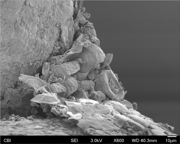
I would like to know more about what exactly is going on in this image, but it seems to show signs of having been separated (or torn, cut, etc) from other biological material. I feel like this image could be a set piece from some sci-fi movie, too.
Here’s the closest we zoomed in:
We could see individual cells in the quinoa. Pretty awesome.
Here’s a quinoa plant for reference:

Next, in my series of “trendy foods under the microscope” I’ll examine a kale smoothie.
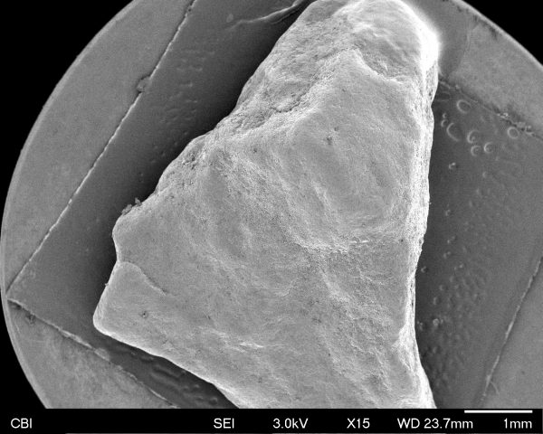
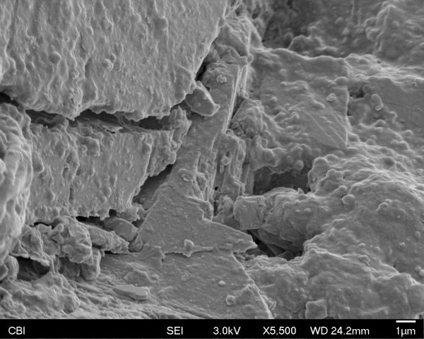
I really enjoyed getting to see things from a view the naked eye could not. I decided to choose something ordinary, because I was curious to see if we could find interesting figures behind very common objects. So I chose a piece of rock I picked up on Forbes near Craig Street. It was interesting to find that the rock had both very rectangular structures and very round structures like the 2 pictures shown below. I also found it very interesting to find different “landscapes” on the surface of the rock. Overall, I was very happy with my results. Hopefully, I will have an opportunity next time to bring in even more interesting objects to see!

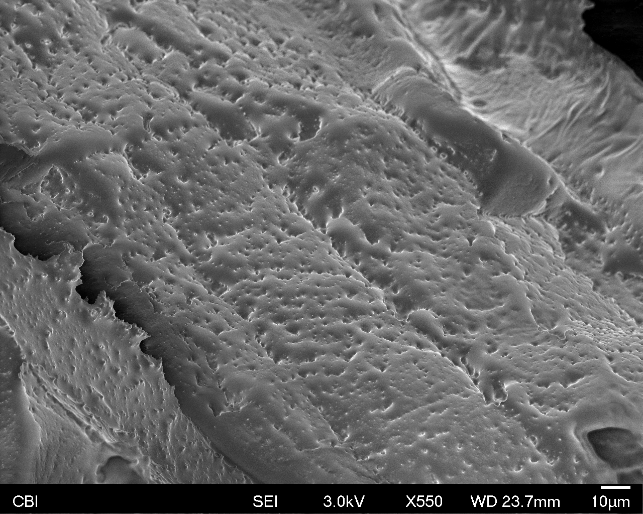
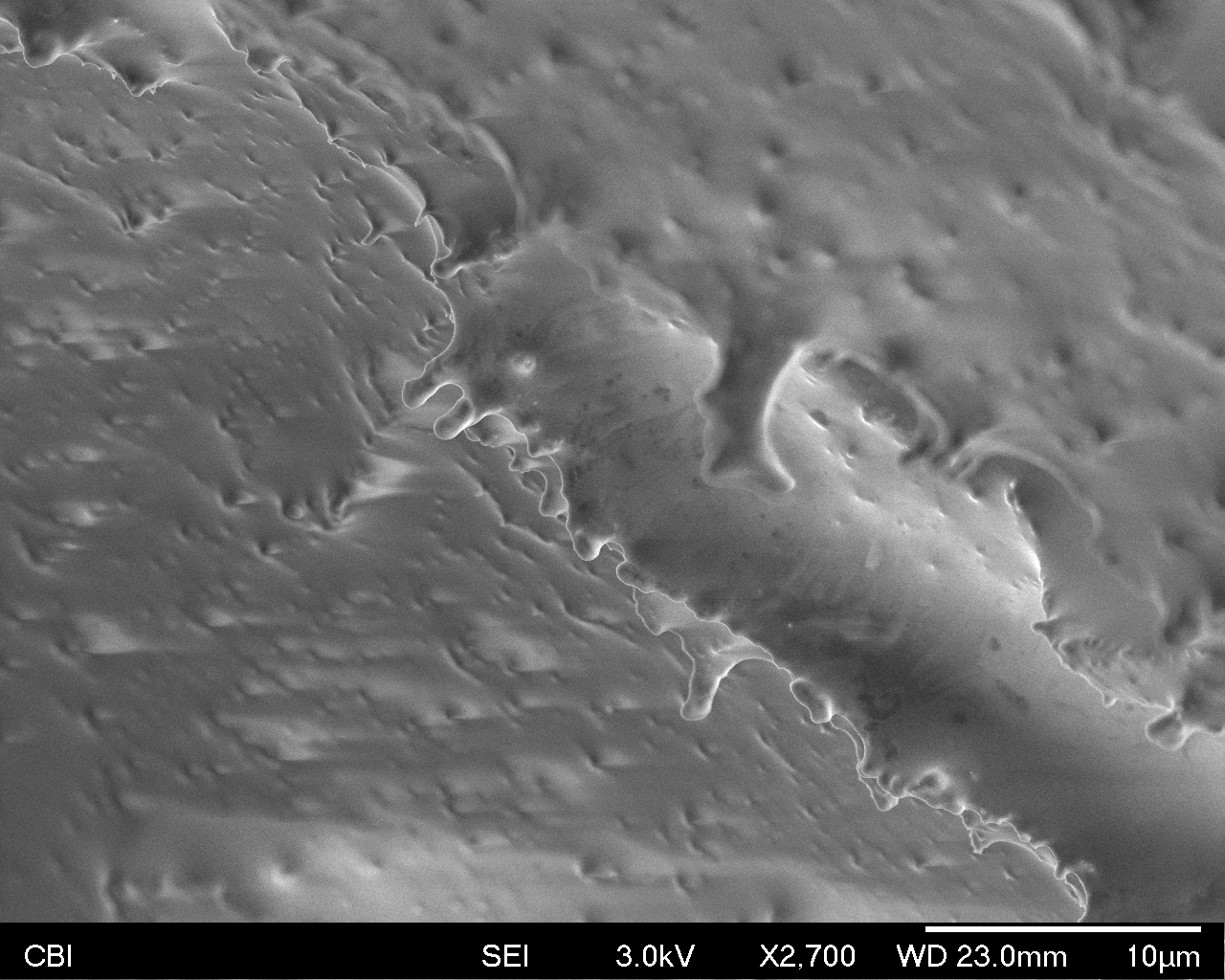
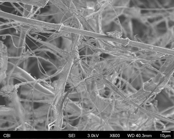
For my SEM imaging, I chose to a piece of twig to scan. At first, I was a bit worried that my “thing” was too boring. This was what it looked like at a recognizable scale. It looked like a monochromatic hot pocket.
Here was a side view of the twig, where I tore it off when snapping the piece from the rest of the twig. Looking at the twig zoomed in and seeing its chaotic fibers was actually really surprising, since I really took for granted what kind of chaos and entropy I could cause as a human onto these small objects.
This was another side view of one of the veins inside the branch. The structure and repeated pattern of the pores was really interesting to me.
Before having read the excerpt from Robert Hooke’s Micrographia, I thought a cork would be an interesting object to look at on a microscopic level due to it’s unique consistency. A cork definitely has some give when you squeeze it, and you can tell that it must be a relatively porous material. The images are in order from furthest away to closest up.
My favorite two observations from these images are first: examining the walls between the cells inside the cork. I thought it was really interesting that there seems to be some sort of material keeping the cells adhered to each other. Second, I enjoyed the last image in the series that shows one of the cell walls. I asked Donna what it was, and she said that it was probably the walls cracking from dehydration.
I had a bouquet of dried roses at home and ripped a small piece from its petal to scan it.
At first, it resembled of a terrain of some planet. We could see where the cracks were and see what’s inside on the edges.
It was a pleasant surprise to discover that cell of dried rose petal actually look like one. The shot above is where the petal’s surface meets the crack, where the interior is revealed. It looks like a pile of leaves on the left, while small cells are tangled tightly together on the other half.
How mesmerizing it is to see that appearance of a cell actually resembles its entity! These shots portray so much about roses’ personality: delicate, slender and exquisite.
This shot also reminded me of dried fruits.

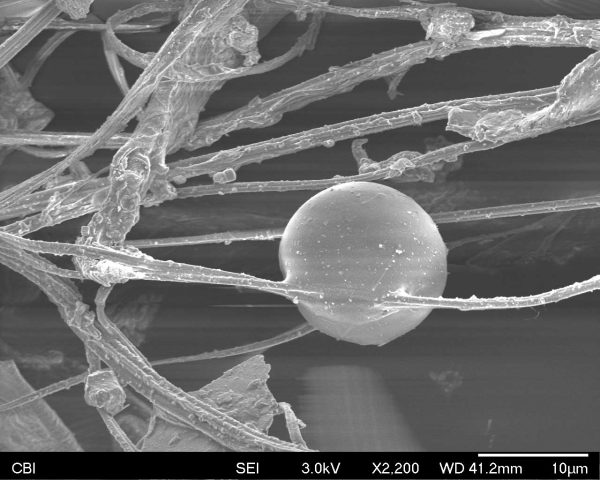
Last Friday I found some wacky stuff in a piece of Ibuprofen.
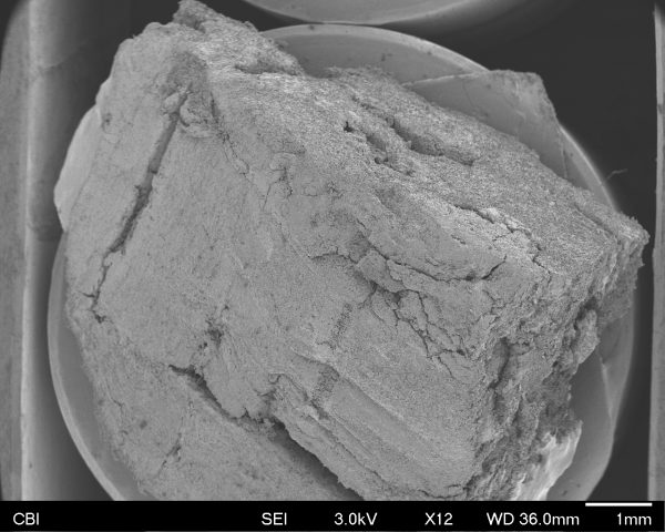
Here’s the Ibuprofen tablet at a recognizable distance: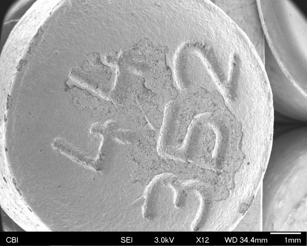
Here it is way zoomed in on some of the chipped off area: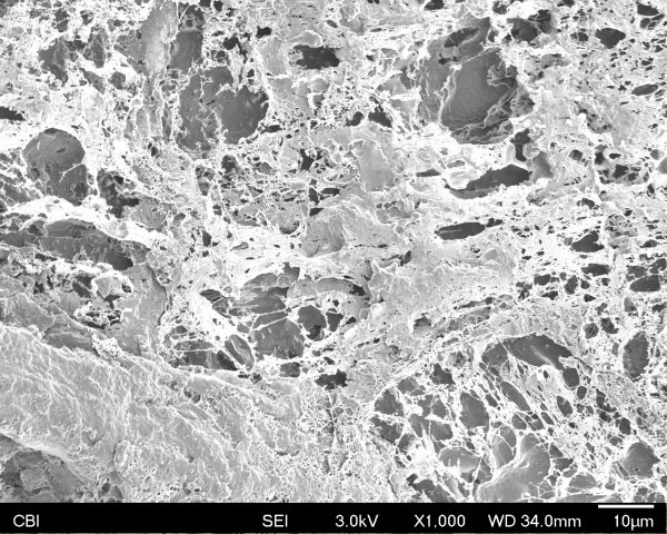
Here it is zoomed in on some cool alien planet lump: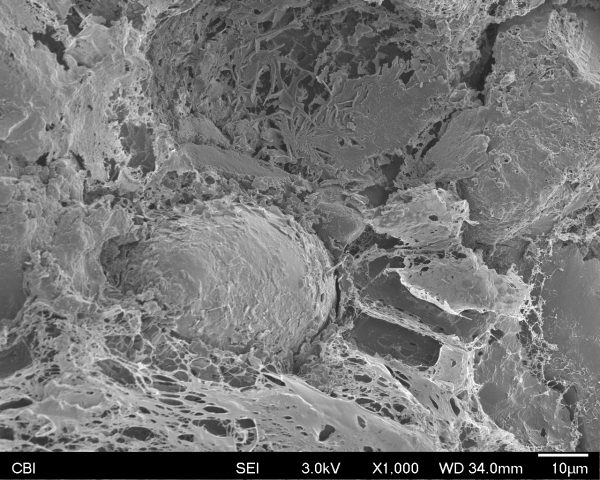
This morning I took an Ibuprofen because my stomach hurt and I just stared at it for so long because now I know that there are entire worlds contained inside this tiny thing I’m about to eat.
I was originally worried that my sample would look really boring, because of the coating that companies put on pills. However, since the pill had been rattling around in my backpack for so long, the coating had worn off in places. The really interesting images came from zooming in on the spots where the coating had worn off. There were a few other locations with tiny bits of the coating chipped off where you could see some of the cool spider-webby stuff hidden just behind a crack in the smooth surface.
After reading Robert Hooke’s, Micrographia, I was really excited to see what he was talking about. Actually using the electron microscope was quite an experience. I honestly didn’t really know what to expect at all. I didn’t know anything about the lab, what the machine was, and what my little piece of cork would look like at a micro scale. So everything was a great surprise. The entire time I kept forgetting how small my piece of cork was and my sense of scale was constantly screwing me over. It was so odd to zoom so far in live. What we saw was pretty fantastic. The natural shapes and curves were beautiful and hard to believe. The whole experience was just unforgettable and I am still amazed that we have the technology to be able to observe anything in that scale.
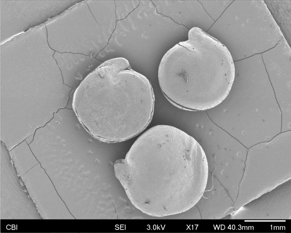
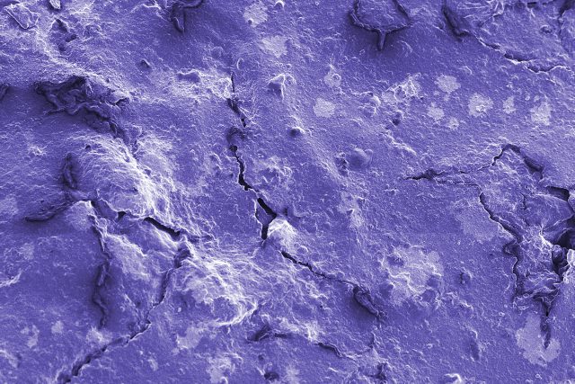
I scanned a little, purple rock picked up from the dirt of a potted plant within Zebra Lounge in the CFA. I learned quite a few things during the scanning. For example, slightest vibrations in the building itself can blur or skew scans! Also, in addition to the scanner using electrons rather than optical imaging, the reason why something at this scale has no color is because it is smaller than colored light itself! For example, purple (the “smallest color” in terms of wavelength, has a wavelength of 400nm, while our scans could be as little as 10nm! I also learned that she needs to add a special liquid to function as a “ground” for the electrons that don’t bounce pack, to prevent static/unintended “light” in the images.
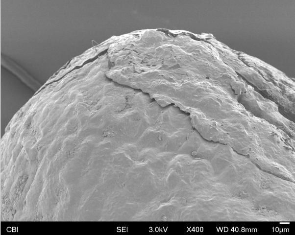
As I scanned the rock in the lab, we found a lot of interesting little things on it. Aside from the expected cracked paint, we found what could be carbonate crystals, possibly from the plant itself. The image above is an example of the carbonate build-up on top of the paint, and below is a close-up view of the crystals.
and it looks like we also found some bacteria colonies (below). The left shows a possible mucus excreted by the bacteria to “contain” them in a moist environment.
Here is a view of the paint cracks:
Here is the edge of the rock, near the top where there may be carbon buildup. It creates a sort of landscape:
And a zoomed-out view:
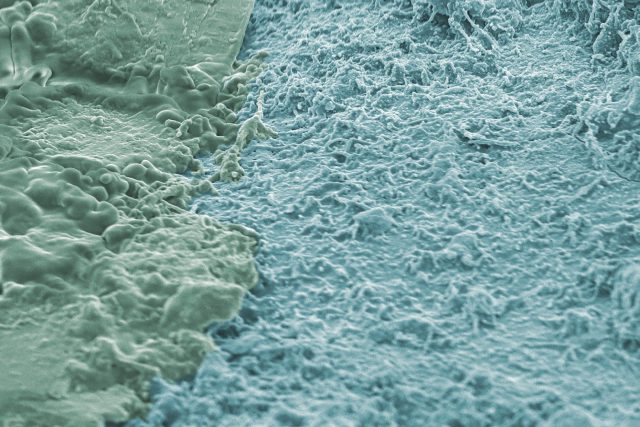
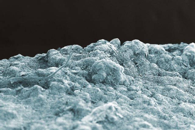
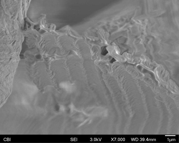
I’ve had my pet Mali Uromastyx lizard, Typhius, for about ten years now. Uromastyx lizards may be mostly drab in color, but they certainly have interesting scales, so scanning some shed skin from my pet felt like a natural choice for the SEM. 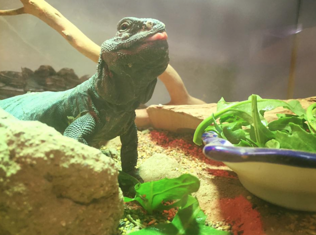
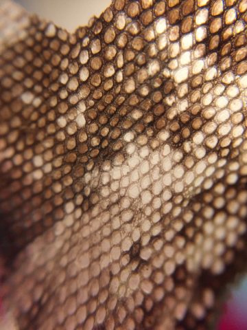
Even under a cheap cell phone macro lens, the scales look pretty neat:
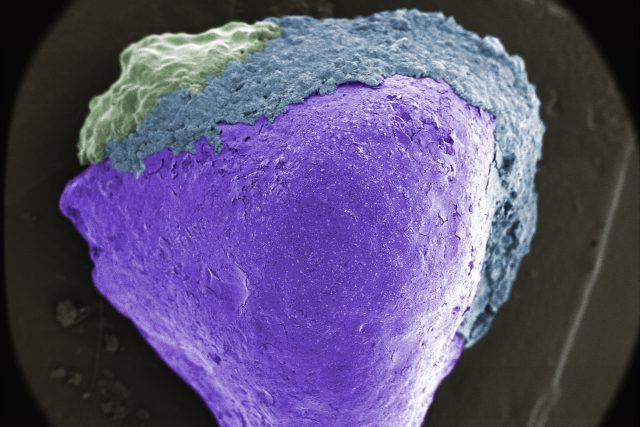
For the sample, I chose a section of skin from near the base of Typhius’s tail. The scales there were pointy looking, but small enough that I had a hard time really examining their geometry. Even at the lowest magnification, in the first picture, the forms looked incredible and revealed some hidden features between the large scales; moving in to 80X and the minuscule surface texture of the scales themselves began to reveal itself!
An interesting feature of this second image is that there seems to be a “light” illuminating the inner geometries. Donna said this was probably due to the electron beam being caught bouncing around under some of the scales, thus increasing the exposure on the sensor and creating the illusion of an extra light source.
The third image shows the highest level of magnification that we looked at the scales under- 450X. At this level, the tiniest surface textures become jagged and rough, going so far as to even reveal the edges of the individual cells! How incredible- this tiny piece of shed skin has revealed itself as an immense alien landscape.

When I tell people what I study, I am usually quickly interrupted with the question “How on earth do neuroscience and art overlap?” Depending on my interlocutor, I don’t always know what to answer. This here is exactly what I wish I could show them. These following images were made with a piece of dead butterfly wing. I remember the way my mom rolled her eyes when I asked her to rummage through her purse so I could use her tampon box as a dead butterfly container. She wasn’t too happy about it. I can’t wait to show these to her now.
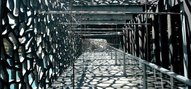
The most interesting part of my session with Donna was noticing how incredibly structural the wings were. They are extremely delicate, to the point where they dissolve between your fingers, but due to their incredible architecture they are able to be used as a reliable, energetic and strong mode of transportation. I found the symmetry and intricacy of the wings absolutely fascinating. At the bottom I attached a picture of the MuCEM (Musee des Civilisations), whose architecture gets pretty close to the microscopic detail of the butterfly wing .
I chose a square of a holographic book cover to scan. When under slight magnification, it quickly became clear that this square was extremely smooth
While I was hoping to find the microridges that makes the hologram effect on many book covers, these ridges are covered by a transparent polymer coating. However, it is only transparent to light, and the electrons bounced right off it without revealing any of the forms below. The coating is smooth and relatively featureless. We had to zoom in until the sharp edges of the square became a rolling incline before any distinct surface features were visible.
Looks a lot like Enceladus, Saturn’s icy satellite. However, instead of hosting subterranean ocean underneath, the coating gives way to the tangled fibers of paper.
Also visible are sections where the coating has chipped off, revealing the tiny holographic grid beneath (center right). We tried to zoom in on that section, but couldn’t get a clean resolution before our time was up.
Color film is made in layers of emulsions and filters. One way to think about it is three layers of black and white film that are in layers, and each layer is sensitive to different color channels. Usually, 3 layers: yellow, magenta, and cyan on the film base, but some films have as much as 12 – and the Ferraria revival project is making a color film with 16 layers.
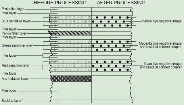
I was never quite satisfied with diagrams like the above. Film isn’t that thick! What’s really going on? To photograph the actual silver halide sensitive-to-light grains, one would need to strip the protective layers off the top with chemical baths. I couldn’t do this, but examined film anyway to see what I would end up with.
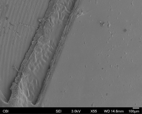
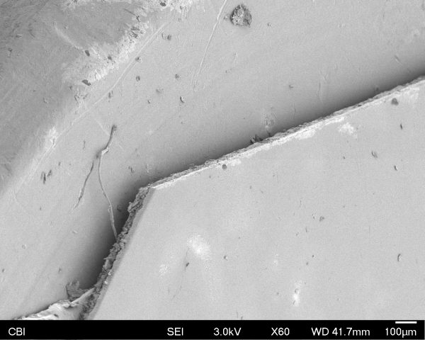
On the left, you can see the metal slug. To the right of that, you can see a bit of the copper tape, and the solver contact in the bottom left corner. The sheet that takes up more than half of the frame, on the right, is the film. Little bits of what is most likely dust on top of the smooth layer. The edge is where the action is however.
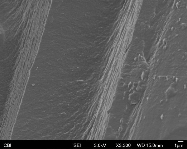
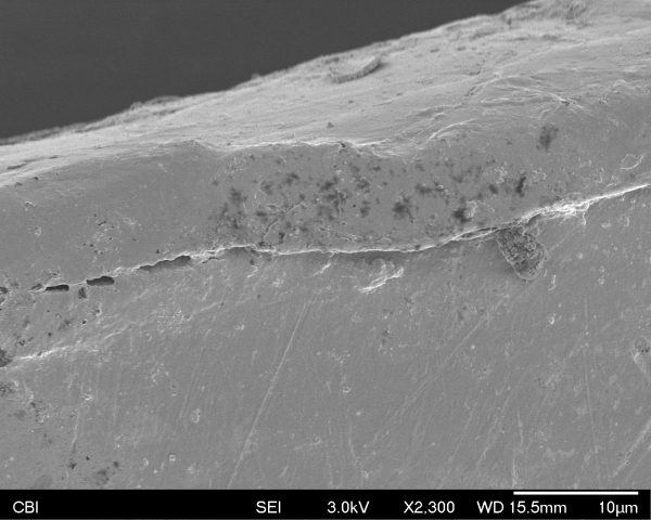
A close up of the edge shows us distinct “shelves”, the different layers of the film, certainly!
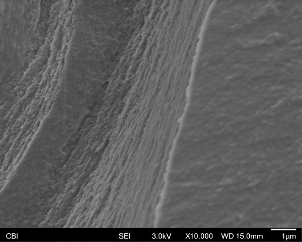
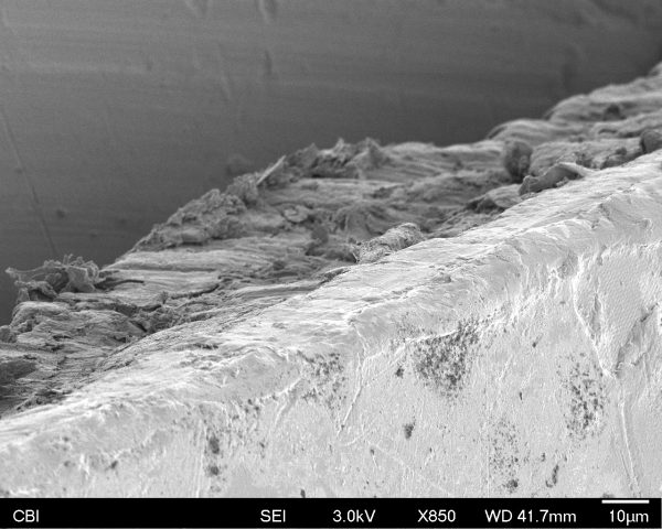
Each of these layers has an interesting amount of texture to them. Perhaps from the crystalline inner structure. Below shows an even closer look.
Ultimately the inner dynamics of film are no more revealed to me than they were before, but seeing it for myself – in real life – is far more reassuring and gratifying than through diagrams or drawings.
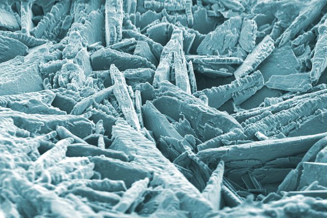
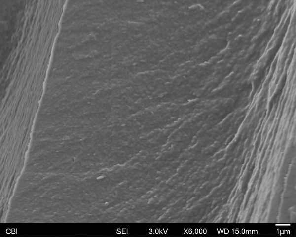
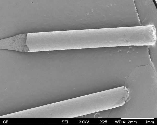
I chose to examine closely a piece of pencil lead:
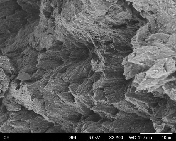
When Donna started operating the joystick for the first time, I instantly felt as though she was operating a rover that was charting a new world. It was so interesting how zooming in far enough completely erases all sense of familiarity, and establishes a whole new terrain void of all sense of scale.
This is an example of that unfamiliarity of scale. At one point, I thought I was seeing a beach within a piece of pencil lead. If not for the information displayed on the bottom and my crude colorization, I wonder if people would be able to tell.
Overall, I think there are great parallels and contrasts to be drawn from seeing the extremes of the minuscule and the massive. I noticed that I felt a similar sense of nebulous beauty from the SEM experience as I do when I go to the planetarium or look up with a telescope. However, I think there is a great contrast in that understanding the vastness of space could make one feel absolutely insignificant, while the SEM images might make that same person feel as if they are the center of the universe.
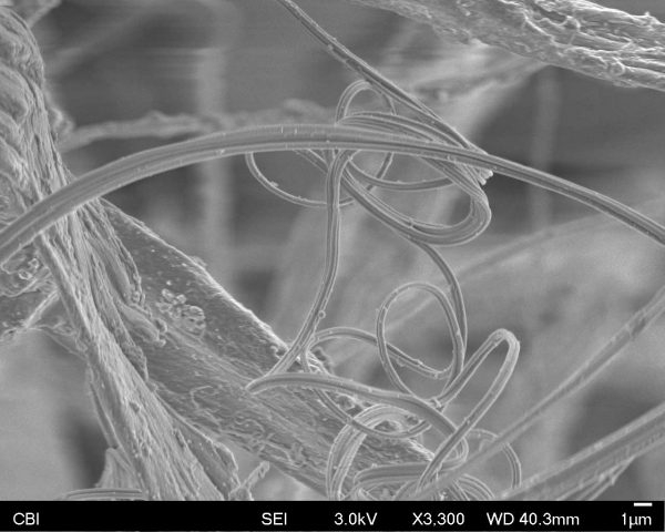
The spider web I scanned had been staying for a while above one of bathtubs in my house: it was most probably not sheer silk anymore. But who would have thought that this whole mess of dust, and who knows what else, would have produced such images. Our brain is not ready for that. It starts comparing what we see to familiar environment: plants, bamboos and barb-wire, bones etc… But this is not it: the very small does exist, and we just have no clue about its beauty, its intricacies and patterns. Nature’s creativity is endless, and while there is a lot to study at the human scale, there is more inspiration to be found for scientists and artists at the scale even below!
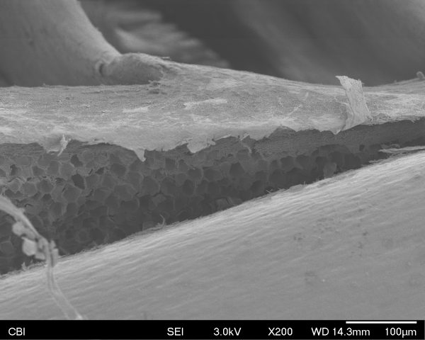
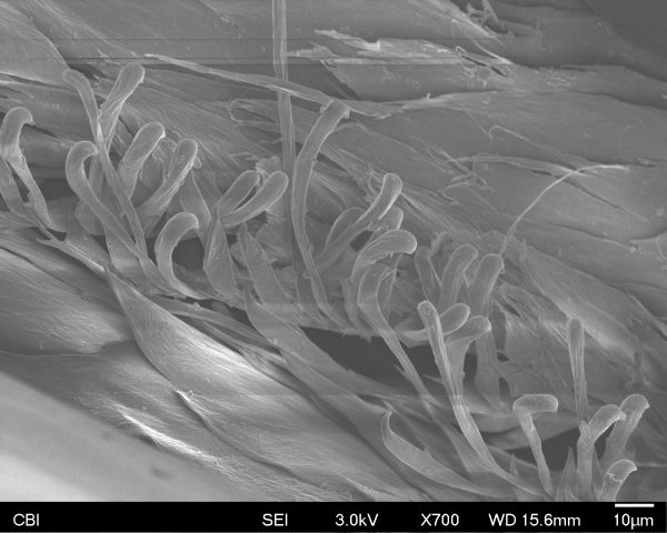
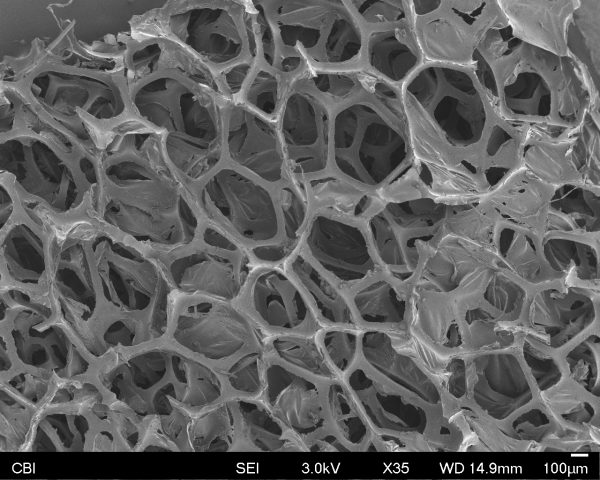
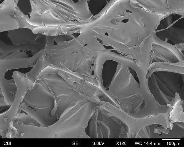
I chose to examine a feather and some very thin packaging foam.
This is the feather:
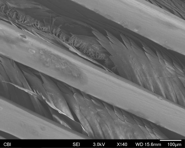
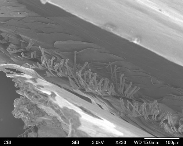
As we increase the magnification, we can see hooks on the feather which keep the tiny fibers aligned. The hooks are sort of mussed up here.
I also examined some packing foam:
During the trip to the SEM lab, I learned that the lens was actually perpendicular to the image plane. Elections would be scattered in all directions off of the object and then could be recorded from any angle. I am curious as to how these images can be converted back into 3d data– as in a world that one could walk through in Unity. That was the compulsion of everyone who saw the SEM images. They had such a foreign texture and such interesting crevices (more like rooms). I would like to see them from a more angles… and magnified to a room-size scale. The items I picked were also really great for such an endeavor as they were not flat but filled with pockets to explore.
http://gifmaker.cc/PlayGIFAnimation.php?folder=2017020705lVRutf9mlMWXs2FNDqTnD0&file=output_ab3OCJ.gif